Silver, BiTe and Antimonene Based Surface Plasmon Resonance Sensor for Enhancement of Sensitivity
Arun Uniyal1,*, Brajlata Chauhan1, Amrindra Pal1, Sanjiv Tomar1 and Priyanka Dobhal Uniyal2
1Department of ECE, School of Engineering and Technology, DIT University, Dehradun, Uttarakhand, India-248009
2GIC Nagnath Pokhari, Chamoli, Uttarakhand, India
E-mail: arunavenue@gmail.com
*Corresponding Author
Received 19 April 2023; Accepted 01 June 2023; Publication 31 July 2023
Abstract
A biosensor based on the surface plasmon resonance has been theoretically analyzed. The proposed surface plasmon resonance (SPR) sensor consists of a hybrid structure with Ag/Bismuth telluride (BiTe)/Antimonene (An)/Sensing medium layers. BK7 is used as the main coupling prism. The sensor’s sensitivity performance gets enhanced by depositing BiTe and antimonene layers over the conventional sensor’s configuration. The Ag, BiTe, and antimonene layer optimized thickness were taken as 45 nm, 0.5 nm, and 0.5 nm, respectively. The proposed sensor has been working on attenuated total reflection. The proposed sensor shows the highest sensitivity of 154 degree/RIU, a signal-to-noise ratio of 0.19 degree with a figure of merit of 29.26 RIU. The proposed SPR sensor can be used for bio analyte and biochemical detection.
Keywords: SPR, sensitivity, Kretschmann, SNR, figure of merit, BK7.
1 Introduction
The surface plasmon resonance optical phenomenon occurs at the metal-dielectric interface. The outcome of this mechanism gives rise to surface plasmons (SPs) which generally arise when free electrons accumulate in the cloud form over a metallic surface. The excitation of plasmons generally depends upon the angle of incidence at which the maximum absorption of incident waves occurs, giving rise to an evanescent wave. The optical biosensors facilitate the real-time monitoring of analytes; no requirement of tags, greater sensitivity, etc., are its merits [1–3]. Fields like medical, chemical, biological, food safety, and others use this optical sensing technology extensively [4–6]. Kretschmann’s configuration is generally employed to design SPR biosensors, and conventional design configuration includes a metal film placed above a glass prism [7]. Silver (Ag) is the most commonly used metal layer and shows a wave spectrum with sharp resonance and higher sensitivity due to an increased bio-molecular absorption rate [8]. Other metals like gold, aluminum, and nickel can also be employed for SPs generations in SPR sensors [9, 10]. Antimonene is a two-dimensional (2D) material with a honeycomb lattice structure. Its optical qualities of greater carrier mobility, suitable bandgap, and higher chemical performance make it suitable for SPR biosensors [11]. In this proposed study, the BiTe material is a topological insulator acting as a basic recognition element (BRE) material. It has greater mobility of carriers (467 cm/V-s). It enhances the sensitivity of the proposed biosensor [12].
A novel SPR design has been proposed in the suggested study, including an Ag-BiTe-Antimonene-Sensing medium. The sensitivity, figure of merit, and signal-to-noise ratio (performance parameters) have been evaluated. Section 2 gives the modeling and analysis of the proposed sensor. Finally, the simulated results have been shown in Section 3.
2 Modelling and Analysis
2.1 Design Parameters
The proposed SPR biosensor comprises five layers, including metal and 2-D materials (Ag An BiTe sensing media). The widths of these layers are nm (Ag), nm (BiTe), nm (Antimonene), and the corresponding refractive index (RI) are , , considered, respectively. Here B and An denotes the number of layers of BiTe and antimonene, respectively. The sensing medium is an aqueous solution (water) of RI 1.33. This RI (n) change is due to the metallic surface’s bio-molecular interaction process (between analytes and immobilized ligands). The RI of the Ag metal is calculated using the Drude-Lorentz model, as given below [13]:
| (1) |
where are collision and plasma wavelength, respectively. Its typical values are and respectively. BK7 is used as a coupling prism. The input TM polarized wave with 633 nm is used in this study. The RI of the prism is 1.515 [14].
2.2 Mathematical Modeling
Mathematical modeling is to be done with the help of the Drude-Lorentz model and the Fresnel equation. The simulation is being carried out in MATLAB 2016a. The reflectance of incident TM p-polarized wave is investigated using an efficient transfer matrix method (TMM) which uses no approximations [15]. The proposed SPR sensor is shown in Figure 1.

Figure 1 Proposed SPR sensor design.
With the z-axis, the layers are layered. The tangent fields at the first and final boundaries are related with the expression [16]:
| (2) |
Where and the electric and magnetic field components are at the first boundary. X and signifies the similar field components for the final boundary. Indicates the characteristic transfer matrix of the sensor design. For TM mode (p-polarized wave), the transfer matrix is expressed as [17]:
| (3) |
The t layer matrix,
| (4) |
denotes the value of optical admittance. It is expressed by:
and phase factor, where and represents incidence angle & wave number in free space. For N – the layer SPR sensor, the value of reflectance is given by [18]:
| (5) |
The performance of an SPR-based biosensor can be analyzed with the help of performance parameters [19]. Firstly, Sensitivity is given by:
Here, shows variation in resonance angle, and shows variation in RI of the sensing medium. The next parameter is signal to noise ratio is generally defined as:
This parameter gives the accuracy of our proposed sensor Figure of merit can be generally expressed as:
The desired values for these parameters are to be on the higher side. The greater the values are, the greater the performance of the proposed sensor.
3 Results and Discussions
Figure 2 gives the reflectivity plots for SPR curves with the angle of incidence for different layer combinations for two different RI of 1.33 and 1.34. It is to be noted that the dip in the SPR curve signifies the minimum reflectance or maximum absorption of light. With the help of Table 1, the values for sensitivity and change in resonance angle () for the four possible cases are shown in Figure 2 [(a)–(d)], and the layer combination case for which maximum sensitivity is achieved (B 1, An 16) is tabulated. As clear from Table 1, the impact of adding both the B and An layers to the conventional SPR sensor design the sensitivity enhances.
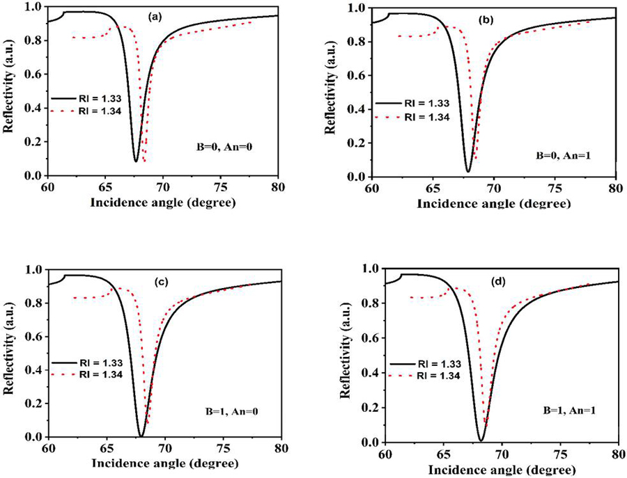
Figure 2 Reflectivity as a function of incidence angle for four different layer combination cases. (a) (B 0, An 0), (b) (B 0, An 1, (c) (B 1, An 0) and (d) (B 1, An 1)).
The analysis made here signifies the impact of RI on the sensing medium.
By increasing the number of layers of antimonene (from 1 to 16) and keeping the BiTe layer constant (B 1), The maximum sensitivity achieved is 154 degrees/RIU. This shows that the sensitivity can be increased to some extent after increasing the value of the layer. After a limit, the sensitivity degrades even while increasing the number of layers keeping another layer constant. This is due to the decrement in the rate of light utilization.
Table 1 Parameters computation for different layer combinations
| Layers | Sensitivity (Degree/RIU) | |
| 116.5 | 1.165 | |
| 117 | 1.17 | |
| 117.8 | 1.178 | |
| 118.1 | 1.181 | |
| 154 (max) | 1.54 |
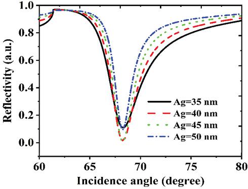
Figure 3 Width optimization curves for metal (Ag) layer.
Figure 3 gives the optimization curves for the silver layer. This width optimization plot shows the minimum reflectance variations for four widths (i.e., 35 nm, 40 nm, 45 nm, and 50 nm). The conclusion made with this plot is that the 45 nm thickness of the Ag layer has minimum reflectance, as minimum reflectance signifies the concept of SPR excitation at the metal-dielectric boundary. So, 45 nm width is the optimized value for the metal (Ag) layer. The next plot (Figure 4) is the outcome of varying a layer taking another layer value as constant. Case 1 gives the impact of increasing the BiTe () layer and taking the monolayer of antimonene (). We see that the SPR curve width increases with the increase in the incident angle. Case 2 impacts reflectivity after varying the antimonene layers and keeping the constant monolayer of BiTe. The trend followed by the SPR curves is almost the same.
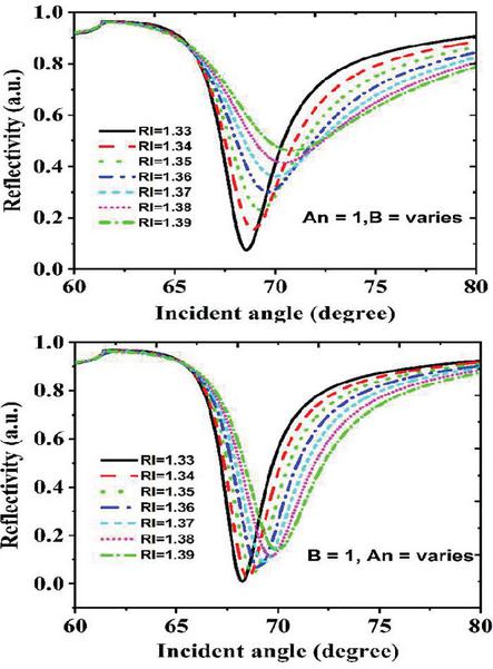
Figure 4 Reflectivity as a function of incidence angle for Case1, and and case 2, and .
4 Comparative Analysis
A comparative analysis of the proposed study with the previous studies has been tabulated with the help of Table 2. Our proposed design’s high sensitivity indicates that our design materials improve the sensitivity of the SPR biosensor. The sensitivity of the proposed sensor attains its maximum value for sensitivity for sixteen antimonene and mono BiTe layers (i.e., B 1, An 16). The other parameters, like FOM, also get enhanced using the proposed structure with 29.26 RIU. However, the value of SNR is in the acceptable range.
Table 2 Proposed and previous SPR designs comparison
| Parameters | ||||
| Ref. | Configuration | S | SNR | FOM |
| This work | Ag/BiTe/An | 154 | 0.19 | 29.26 |
| [20] | Ag/graphene | 91.76 | – | – |
| [21] | Ag/graphene/MoS/TiO/SiO | 98 | 0.88 | 87.11 |
| [22] | ZnO/Ag/Au/BaTiO/ Graphene | 116.67 | 4.54 | 37.87 |
5 Conclusion
The modified Kretschmann’s configuration uses prism coupling for the SPR biosensor design. The optimized thickness of Ag is taken as 45 nm with 0.5 nm of both BiTe and antimonene. The maximum sensitivity attained here is 154 degree/RIU, FOM of 29.26 RIU and a signal-to-noise ratio of 0.19 degree has been achieved. The future scope of this proposed four-layer sensor design includes its application in various disease diagnoses like malaria, cancer, etc., or maybe in detecting different types of bacteria, viruses, etc.
Acknowledgments
We thank DIT University for its continuous support of this research work.
References
[1] J. Homola, “Present and future of surface plasmon resonance biosensors,” Anal. Bioanal. Chem., vol. 377, no. 3, pp. 528–539, 2003, doi: 10.1007/s00216-003-2101-0.
[2] P. Damborský, J. Švitel, and J. Katrlík, “Optical biosensors,” Essays Biochem., vol. 60, no. 1, pp. 91–100, 2016, doi: 10.1042/EBC20150010.
[3] B. Karki, A. Uniyal, A. Pal, and V. Srivastava, “Advances in Surface Plasmon Resonance-Based Biosensor Technologies for Cancer Cell Detection,” Int. J. Opt., vol. 2022, 2022, doi: 10.1016/j.bios.2021.113767.
[4] J. Zhang, L. Zhang, and W. Xu, “Surface plasmon polaritons: Physics and applications,” J. Phys. D. Appl. Phys., vol. 45, no. 11, 2012, doi: 10.1088/0022-3727/45/11/113001.
[5] Y. Kumar, R. Mishra, E. Panwar, J. Kaur, and R. Panwar, “Design, optimization and critical analysis of graphene based surface plasmon resonance sensor for DNA hybridization,” Opt. Quantum Electron., vol. 51, no. 10, 2019, doi: 10.1007/s11082-019-2057-8.
[6] A. Uniyal, G. Srivastava, A. Pal, S. Taya, and A. Muduli, “Recent Advances in Optical Biosensors for Sensing Applications: a Review,” Plasmonics, no. 0123456789, 2023, doi: 10.1007/s11468-023-01803-2.
[7] E. Kretschmann and H. Raether, “Radiative decay of non-radiative surface plasmons by light,” Z. Naturforsch, vol. 23, no. a, pp. 2135–2136, 1968.
[8] R. Kashyap et al., “Enhanced biosensing activity of bimetallic surface plasmon resonance sensor,” Photonics, vol. 6, no. 4, 2019, doi: 10.3390/photonics6040108.
[9] A. Nisha, P. Maheswari, P. M. Anbarasan, K. B. Rajesh, and Z. Jaroszewicz, “Sensitivity enhancement of surface plasmon resonance sensor with 2D material covered noble and magnetic material (Ni),” Opt. Quantum Electron., vol. 51, no. 1, 2019, doi: 10.1007/s11082-018-1726-3.
[10] A. H. M. Almawgani, P. Sarkar, A. Pal, G. Srivastava, and A. Uniyal, “Titanium Disilicide, Black Phosphorus – Based Surface Plasmon Resonance Sensor for Dengue Detection,” Plasmonics, no. 0123456789, 2023, doi: 10.1007/s11468-023-01856-3.
[11] A. Uniyal, B. Chauhan, A. Pal, and Y. Singh, “Surface plasmon biosensor based on BiTe antimonene heterostructure for the detection of cancer cells,” Appl. Opt., vol. 61, no. 13, pp. 3711–3719, 2022.
[12] Y. Zhao, S. Gan, G. Zhang, and X. Dai, “High sensitivity refractive index sensor based on surface plasmon resonance with topological insulator,” Results Phys., vol. 14, no. June, p. 102477, 2019, doi: 10.1016/j.rinp.2019.102477.
[13] A. Uniyal, S. Gotam, T. Ram, B. Chauhan, A. Jha, A. Pal. (2022), “Next Generation Ultra-sensitive Surface Plasmon Resonance Biosensors”. In: Khare, N., Tomar, D.S., Ahirwal, M.K., Semwal, V.B., Soni, V. (eds) Machine Learning, Image Processing, Network Security and Data Sciences. MIND 2022. Communications in Computer and Information Science, vol 1762, 2022. Springer, Cham. https://doi.org/10.1007/978-3-031-24352-3\_31.
[14] A. Uniyal, A. Pal, and B. Chauhan, “Long-Range Spr Sensor Employing Platinum Diselenide and Cytop Nanolayers Giving Improved Performance,” Phys. B Condens. Matter, vol. 649, no. September 2022, p. 414487, 2022, doi: 10.2139/ssrn.4230023.
[15] B. Karki, A. Uniyal, G. Srivastava, and A. Pal, “Black Phosphorous and Cytop Nanofilm-Based Long-Range SPR Sensor with Enhanced Quality Factor,” J. Sensors, vol. 2023, 2023, doi: 10.1155/2023/2102915.
[16] Y. Singh and S. K. Raghuwanshi, “Titanium dioxide (TiO) coated optical fiber-based SPR sensor in near-infrared region with bimetallic structure for enhanced sensitivity,” Optik (Stuttg)., vol. 226, no. P1, p. 165842, 2021, doi: 10.1016/j.ijleo.2020.165842.
[17] S. Singh, A. K. Sharma, P. Lohia, D. K. Dwivedi, V. Kumar, and P. K. Singh, “Simulation study of reconfigurable surface plasmon resonance refractive index sensor employing bismuth telluride and MXene nanomaterial for cancer cell detection,” Phys. Scr., vol. 98, no. 2, 2023, doi: 10.1088/1402-4896/acb023.
[18] B. Karki, G. Ansari, A. Uniyal, and V. Srivastava, “PtSe2 and black phosphorus employed for sensitivity improvement in the surface plasmon resonance sensor,” J. Comput. Electron., no. 0123456789, 2022, doi: 10.1007/s10825-022-01975-w.
[19] N. Mudgal, A. Saharia, A. Agarwal, J. Ali, P. Yupapin, and G. Singh, “Modeling of highly sensitive surface plasmon resonance (SPR) sensor for urine glucose detection,” Opt. Quantum Electron., vol. 52, no. 6, pp. 1–14, 2020, doi: 10.1007/s11082-020-02427-0.
[20] P. K. Maharana, P. Padhy, and R. Jha, “On the Field Enhancement and Performance of an Ultra-Stable SPR Biosensor Based on Graphene,” IEEE PHOTONICS Technol. Lett., vol. 25, no. 22, pp. 2156–2159, 2013.
[21] M. Moznuzzaman, M. Rafiqul Islam, M. Biplob Hossain, and I. Mustafa Mehedi, “Modeling of highly improved SPR sensor for formalin detection,” Results Phys., vol. 16, no. September 2019, p. 102874, 2020, doi: 10.1016/j.rinp.2019.102874.
[22] N. Mudgal, A. Saharia, A. Agarwal, and G. Singh, “ZnO and Bi-metallic (Ag–Au) Layers Based Surface Plasmon Resonance (SPR) Biosensor with BaTiO and Graphene for Biosensing Applications,” IETE J. Res., 2020, doi: 10.1080/03772063.2020.1844074.
Biographies

Arun Uniyal received his Master’s degree in Wireless Mobile Communication from Uttarakhand Technical University, Dehradun, India. Currently, he is working as an Assistant Professor (ECED) at Institute of Technology, Gopeshwar, India. Also persuing PhD from DIT University, Dehradun, Uttarakhand,India. His current area of interest is Optical biosensors, Plasmonics. He has published more than 15 research articles in his area of interest. In addition, he is instrumental in various other research related activities like reviewing for various reputed journals, organizing/participating in conferences.

Brajlata Chauhan received her Ph.D in Microstrip Conformal Antenna and M.Tech in Digital communication from Uttarakhand Technical University, Dehradun, UK, India, in 2017 & 2010, respectively. She is working as an Assistant Professor in the Department of EECE at DIT University Dehradun, India. She has published more than 35 research papers, three book chapters in the field of MW & Antenna, Photonics, and biosensors in international Journals/conferences, and guided 4 M.Tech students. She is a member of IETE, IEEE, ISTE and has 16 yrs of teaching and research experience.

Amrindra Pal has completed his B. Tech (ECE) from Uttar Pradesh Technical University, Lucknow in 2008, M.E (Electronics Instrumentation and Control) from Thapar University, Patiala in the year 2013, and Ph. D in the field of Design and Modelling of Opto-Electronics Devices from DIT University, Dehradun in the year 2018. He has about ten years of teaching experience at various levels and presently working as Assistant Professor in the School of Engineering and Technology, DIT University, Dehradun, India. He has published 50 papers in International Journals and International Conferences, and patents. His research interest includes Integrated Optics, Plasmonics, Opto-Electronics Devices, Electro-Optics, Optical Biosensor, and RF & Microwave. He has been serving as an editorial review board member for peer-reviewed journals.

Sanjiv Tomar (Retd) has an illustrious career spanning over three decades in the Army. He was commissioned in the Corps of Electronics and Mechanical Engineers and has served across the length and breadth of the country ensuring operational readiness of electronics and communication systems in various capacities. He has a BTech (Electronics) and MTech (Electronics) Degree from Jawahar Lal Nehru University, New Delhi. He earned his Ph.D in Development of UWB Antennas for Indoor Wireless Communication System from Manipal University, Jaipur. He has been a Research Fellow at world renowned Think Tank – Institute for Defence Studies and Analyses, New Delhi and conducted research on Military Applications of Nanotechnology. He has been the Project Leader for execution of a multi-disciplinary weapon system of Indian Army. He has authored a number of research papers which have been published in peer reviewed International Journals.

Priyanka Dobhal Uniyal has completed her Master’s in Mathematics from H.N.B.G.U. Srinagar Uttarakhand in 2014. Presently she has been working as a lecturer in Government Inter College, Nagnath Pokhri, Chamoli, Uttarakhand, India. She was a university topper in M.Sc. (Mathematics) in the year 2014. Her research area includes Numerical Analysis, Mathematical modelling.
Journal of Graphic Era University, Vol. 11_2, 177–190.
doi: 10.13052/jgeu0975-1416.1124
© 2022 River Publishers